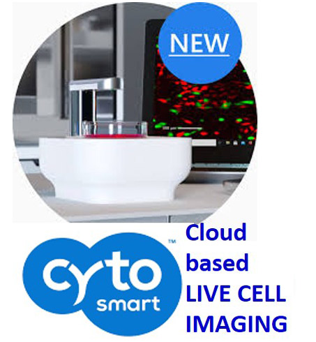
OMNI - The smallest automated live-cell imager that fits within any incubator
The all-new CytoSMART™ Omni is the first affordable automated live-cell imager that fits inside a standard cell culture incubator. It can acquire a bright-field scan of any cell culture vessel within minutes and provides almost instant cloud-based image analysis of your cell culture.
The CytoSMART™ Omni is:
• Fast - Faster than any automated live-cell imager ever seen
• Flexible - Any type of transparent culture vessel can be scanned
• Compact - Fits in any cell culture incubator
The world’s fastest live-cell imager - The CytoSMART™ Omni can acquire a brightfield scan of your entire cell culture vessel within minutes. Not only the scanning process is fast, setting up your experiment is also done within a few minutes, after which the CytoSMART™ Omni does the work for you. The scan is made and uploaded to the CytoSMART™ Cloud (powered by Microsoft Azure) where our custom image analysis algorithms evaluate the scan within minutes. This allows you to quickly set up your experiment and walk away, so you can spend your precious time on data analysis instead of obtaining data.
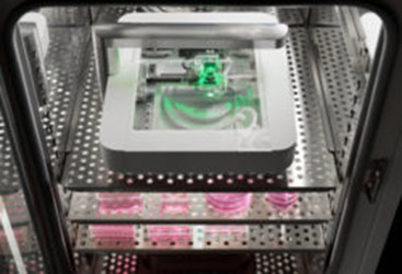
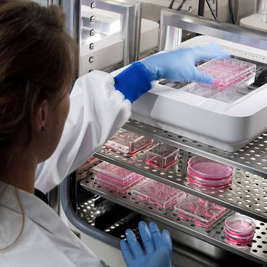
Optimum culture conditions - The CytoSMART™ Omni is so compact, it easily fits inside any cell culture incubator. Next to this, the shape of the CytoSMART™ Omni is especially designed so it does not affect the temperature and airflow inside the incubator. This enables you to perform your experiments at the optimum culture conditions for your cells, without any fluctuations in temperature or CO2-level. Because the CytoSMART™ Omni is placed inside the incubator, you can even scan samples cultured under hypoxia without the need to worry about changes in O2-level. Your colleagues will like this device too, since it is so compact that there is still space left in the incubator for their cell cultures.
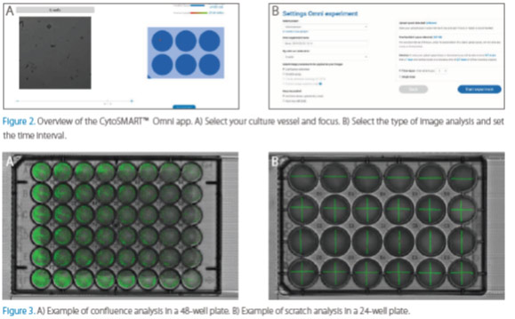
Set up and walk away - The straightforward Omni app (Fig. 2) allows anyone in your lab to use the CytoSMART™ Omni without extensive training. Just place the device inside your cell culture incubator, connect it to a computer and start the Omni app. Place your culture vessel on the optical window of the Omni, follow the intuitive steps of the Omni app, start your experiment and walk away.
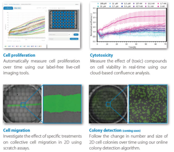
Using the integrated cloud-based image analysis algorithms, you can perform cell culture quality control, proliferation, cytotoxicity and cell migration (wound healing) experiments in just a few clicks. Using commercially available image analysis programs (e.g. ImageJ, Matlab, Python) you can use the images obtained with the CytoSMART™ Omni for applications such as: 3D spheroid formation, neurite formation, angiogenesis, etc